Torso Geometry Reconstruction and Body Surface Electrode Localization Using Three-Dimensional Photography
Abstract
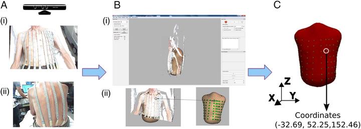
We conducted a prospective clinical study (n=14; 29% female) to assess the accuracy of a three-dimensional (3D) photography-based method of torso geometry reconstruction and body surface electrodes localization. The position of 74 body surface electrocardiographic (ECG) electrodes (diameter 5 mm) was defined by two methods: 3D photography, and CT (marker diameter 2 mm) or MRI (marker size 10×20 mm) imaging. Bland-Altman analysis showed good agreement in X (bias −2.5 [95% limits of agreement (LoA) −19.5 to 14.3] mm), Y (bias −0.1 [95% LoA −14.1 to 13.9] mm), and Z coordinates (bias −0.8 [95% LoA −15.6 to 14.2] mm), as defined by the CT/MRI imaging, and 3D photography. The average Hausdorff distance between the two torso geometry reconstructions was 11.17 ± 3.05 mm. Thus, accurate torso geometry reconstruction using 3D photography is feasible. Body surface ECG electrodes coordinates as defined by the CT/MRI imaging, and 3D photography, are in good agreement.
Jason A. Thomas
PhD
Medical Data & AI Scientist | Strategist | Informatician | Tech lead - Senior Data & AI Scientist - Philips
My research interests include 1) building foundational layers (data, infrastructure, knowledge representation, talent, culture) to support biomedical data science and 2) applying data science & AI methods on data to drive business value and improve patient outcomes.